|
|
Proofs
Introducing new science
History
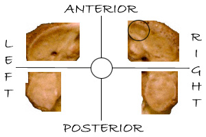
This is a detailed composite showing the superior facets of the human atlas. The right anterior quadrant on this bone shows a divot or erosion due to an incorrect positioning of the head (occipital condyles) over time. This hole is approximately 1/16 inch deep, and can obviously be detected by even a blind man.
Craton's articles on atlanto-occipital dissection (1985)
About the author
See Contract
See also: ATLAS AXIS SKULL O/A JOINTS TEXTBOOK ERRORSERRORS ONLINE:
(1) From
GateWay Community
College
Phoenix, AZ
Created by
Dr. J. Crimando
SUPERIOR VIEW OF THE ATLAS (C1)
(NOTICE SINGULARITY OF SUPERIOR
FACET; THEY SHOULD LIST TWO
FACETS PER SIDE. PLEASE COMPARE.)
(2) From Clinical Imaging
Diagnosis:
DAVID,
atlas of human anatomy
Developed by J. C. Oberson MD.
ATLAS,
SUPERIOR VIEW
Yes, I am fully aware that the three links directly above are no longer working. I do not know for sure that my linking to them is what caused for them to no longer be available. But I do know that it only took about two weeks after I did link to them for their site to disappear. I am hoping that they will be returning in the near future.
| We need your help: | Receive our film | Just give |
This page was first posted on October 21, 2001 and last revised on May 9, 2007.

